Contents
Have you ever wondered why some medical devices fail to capture accurate signals from the body? The issue often isn’t with the device itself—but with the surface it connects to. Yes, we’re talking about surface electrodes. In this article on the Tech4Ultra Electrical website, you’ll discover the critical role they play in measuring bio-electrode potential, and why they are essential components in biomedical instrumentation. Understanding these details can be the difference between accurate readings and misleading data.
What is a Surface Electrode?
I still remember the first time I used a surface electrode in a lab session—it was oddly fascinating. I was expecting something bulky and complex, but instead, it was this simple-looking patch that stuck to the skin. That was the moment I realized how elegant biomedical technology can be. But what really blew my mind was how this tiny thing could detect what’s going on inside your body—without even breaking the skin!
So, what exactly is a surface electrode? It’s a non-invasive sensor that sits on the skin and picks up electrical signals from underneath. Think of it as an eavesdropper that listens to the internal conversations of your body—whether it’s your heart talking through an ECG or your muscles speaking during EMG testing. These electrodes work by capturing the small voltage differences that result from biological activity, known in the field as bio-electrode potential.
Now, if you’re picturing something uncomfortable or hard to use, you’re in for a surprise. Most surface electrodes are made with soft, flexible materials like silver/silver-chloride (Ag/AgCl) and often come with a conductive gel to improve signal quality. You stick them on, record your signals, and peel them off—it’s that simple.
Here are a few key things I learned the hard way, and you should know too:
- Clean skin = better signal: Oils or sweat can mess with the readings big time. Always clean the skin before attaching the electrode.
- Placement matters: Even a small shift can distort your data. You have to be precise.
- Not reusable: Most surface electrodes are designed for single use. Trying to reuse them? Don’t. You’ll get poor data.
There’s a reason why surface electrodes are a cornerstone of biomedical instrumentation. They’re cost-effective, easy to use, and when handled correctly, surprisingly accurate. It’s wild to think that this small patch of material can bridge the gap between our inner bioelectrical world and the screens we use to monitor health.
As the legendary engineer Charles Proteus Steinmetz once said, “There are no foolish questions and no man becomes a fool until he has stopped asking questions.” If you’re diving into the world of biosignals, understanding the surface electrode is the first question you need to answer.
Read Also: Biomedical Instrumentation: Overview, Devices, and Applications
Importance in Biomedical Signal Detection
Contextual Relevance in ECG, EMG, EEG
Back when I was still new to biomedical instrumentation, I underestimated the role of surface electrodes. I figured the machines did all the work. But after struggling with a noisy ECG signal during a demo, I realized something crucial—the quality of your readings starts at the surface. Literally.
Let’s talk about where surface electrodes really shine: in detecting bio-signals like ECG (electrocardiography), EMG (electromyography), and EEG (electroencephalography). These are the bread and butter of non-invasive diagnostics, and the surface electrode is the unsung hero behind them.
- In ECG: These electrodes are placed on the chest or limbs to monitor the heart’s electrical activity. I still remember the first clean ECG trace I got—those perfect little PQRST waves. It felt like I cracked a code. The accuracy of that trace depended entirely on how well those surface patches were placed.
- In EMG: Surface electrodes measure the electrical signals produced by muscles. Whether you’re diagnosing a neuromuscular disorder or analyzing sports performance, these sensors pick up on the smallest muscle contractions. I once tried them during a biceps curl session—it was wild seeing my muscle activity live!
- In EEG: Brainwaves are faint, and surface electrodes on the scalp have to be incredibly sensitive. A friend of mine used to run EEG studies, and he told me stories of losing entire datasets just because an electrode lost contact by a hair. Literally, a hair out of place could ruin it.
The beauty of the surface electrode lies in its versatility. Whether you’re tracking heartbeats, muscle twitches, or brainwaves, it translates the body’s electrical language into readable data. And when you’re dealing with bio-electrode potentials that are only a few millivolts, even the tiniest improvement in contact quality makes a big difference.
Here’s what I learned the hard way:
- Use proper skin prep for better signal quality—especially critical in EEG where the signals are super weak.
- Don’t move during recordings. Even subtle shifts can generate motion artifacts that distort readings.
- In long sessions, watch out for gel drying. It’s a sneaky cause of signal loss.
Simply put, if you’re working in the field of biomedical instrumentation, mastering the use of surface electrodes is non-negotiable. They’re not just tools—they’re your connection to the living data within the human body.
How Surface Electrodes Work
Signal Generation and Bio-Electrode Potential
I used to think electrodes just “picked up signals” like magic. But nope—it’s all about bio-electrode potential. Here’s the gist: when your body generates electrical activity—like from your heart or brain—it creates tiny voltage differences. These are bio-potentials, and surface electrodes detect them by sitting on the skin and catching the electrical changes that reach the surface.
But those signals aren’t floating in the air. They’re traveling through fluids and tissues until they reach the electrode, and that’s where things get interesting (and sometimes messy).
Electrode-Tissue Interface Mechanics
The point where the electrode touches your skin is more than just contact—it’s a micro-environment. I learned this the hard way during a sweaty summer session where signal quality dropped like crazy. The electrode-tissue interface involves complex ion exchange between the skin, the conductive gel, and the electrode material (usually Ag/AgCl). If that balance gets thrown off—say, by dried gel or dirty skin—your signals get noisy, or worse, completely lost.
This interface also introduces impedance, which can mess with the accuracy of the signal. That’s why prepping the skin is just as important as choosing the right electrode.
Skin Surface Detection vs Invasive Techniques
Some people think surface detection is less “reliable” than invasive methods. And sure, invasive techniques—like needle electrodes—do provide super clean signals because they’re right in the tissue. But here’s the thing: surface methods are non-invasive, painless, and safer. No risk of infection, no recovery time. Plus, with today’s tech, surface electrodes can give surprisingly accurate data when used correctly.
Bottom line? Understanding how surface electrodes work gives you power over your measurements. Ignore the details, and you’re just guessing with expensive gear.
Watch Also: Voltaic Cell: Definition, Working, and Examples
Key Components and Materials
Silver/Silver-Chloride Composition
The first time I looked closely at a surface electrode, I was surprised to find out it wasn’t just a piece of foil and tape. The real magic? Silver/silver-chloride (Ag/AgCl). This combo is the industry standard for a reason—it’s stable, non-polarizable, and provides excellent signal quality. When I tried electrodes made with different materials, I noticed more noise and drift. Trust me, stick with Ag/AgCl if you want reliable readings.
Conductive Gels and Adhesives
Another game-changer is the conductive gel. I once used an electrode without gel during an EMG session—big mistake. The signal was barely usable. The gel acts as a bridge between the skin and the electrode, reducing impedance and enhancing conductivity. The adhesive part? That’s more than just sticky stuff—it keeps the electrode in place during movement, which is vital in long recordings or athletic settings.
Durability, Biocompatibility, and Safety
Here’s what I didn’t know at first: not all electrodes are skin-friendly. Good surface electrodes are designed to be biocompatible, meaning they won’t irritate the skin or cause allergic reactions. They’re also disposable for hygiene reasons, but some are made for short-term reuse with high durability. Safety-wise, they’re low-voltage, so no shocks—just clean, passive sensing of your body’s electrical rhythms.
Detailed Classification of Surface Electrodes
Floating Electrodes
My first real experience with floating electrodes was during an EEG study. I was skeptical at first—how could something that doesn’t directly touch the skin give clean signals? But these clever designs use a gel-filled cavity between the electrode and the skin, which “floats” the electrode above the surface. The benefit? Reduced motion artifacts. When subjects move, these electrodes stay stable, and the signal stays smooth.
The catch? They’re a bit more complex to apply and usually cost a bit more. But in high-motion environments—like during physical therapy—they’re worth it.
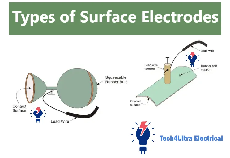
Metal Plate Electrodes
These are the classic types we all probably saw in early physiology classes. Flat, rigid, and made of silver or silver-chloride, metal plate electrodes rest directly on the skin. They’re easy to clean and reuse, which made them popular in labs where cost mattered. I used them a lot in early training because they were durable.
The downside? They’re sensitive to movement and need good skin contact with conductive gel. Without that, you get signal drift or high noise levels. Not ideal for dynamic tests, but great in calm, clinical setups.
Multipoint Electrodes
This design felt futuristic when I first saw it. Multipoint electrodes consist of several small contact points instead of one large surface. The idea is smart: if one point loses contact, others still maintain the signal. I saw them used in EMG applications, where even small muscle movements need precise tracking.
They offer redundancy and better contact over irregular surfaces, but applying them takes longer. Plus, the more components involved, the higher the chance something goes wrong—or breaks.
Comparison Table: Surface Electrode Types
| Type | Advantages | Disadvantages |
|---|---|---|
| Floating Electrodes | Reduced motion artifacts, ideal for movement-heavy studies | More complex setup, higher cost |
| Metal Plate Electrodes | Durable, reusable, cost-effective | Sensitive to movement, requires strong gel contact |
| Multipoint Electrodes | Multiple contact points improve signal reliability | Time-consuming to apply, more components to manage |
Each type of surface electrode has its place, and choosing the right one depends on your specific application—whether it’s clinical, athletic, or research-driven. I’ve tried them all, and trust me, matching the electrode to the context makes a night-and-day difference in your results.
Biomedical Applications
ECG: Heart Monitoring
The first time I used surface electrodes in an ECG setup, I was nervous. Would the heart signals really show up that clearly? Turns out, when you place the electrodes correctly, it’s almost like watching your heartbeat on-screen. That rhythmic pattern—the P wave, QRS complex, T wave—it’s all made visible through these tiny sensors.
Electrocardiography (ECG) is one of the most common uses of surface electrodes. They’re usually placed on the chest and limbs to monitor the electrical activity of the heart. This non-invasive method helps detect arrhythmias, monitor pacemakers, and even assess heart health during physical exertion.
EMG: Muscle Signal Tracking
Electromyography (EMG) changed the way I understood muscles. When I hooked up surface electrodes to my forearm and flexed, I could literally see my muscle activity spike on a graph. It was oddly satisfying—and very eye-opening. These electrodes pick up the tiny voltage changes in muscles during movement, helping in diagnosing neuromuscular disorders or improving rehabilitation programs.
EEG: Brain Activity Detection
Electroencephalography (EEG) is where things get super sensitive. I’ve seen how even minor errors in electrode placement or poor contact can wreck an entire EEG session. In this setup, surface electrodes are placed on the scalp to record brainwave patterns, helping diagnose epilepsy, monitor brain injuries, or even study sleep cycles.
Specialized Use Cases: Sleep Studies and Fitness Wearables
In sleep labs, surface electrodes are indispensable. They help monitor everything from brain activity (EEG) to muscle tone (EMG) and eye movement—all crucial for identifying disorders like sleep apnea or narcolepsy. I once observed a technician prep a patient for a sleep study; by the end of it, they looked like they had antennae—but those signals told an entire story.
Then there’s the modern twist: fitness wearables. From heart rate bands to smart shirts, these gadgets use miniaturized surface electrodes to track your vitals in real-time. Whether you’re a pro athlete or just trying to close your rings, you’ve probably used this tech without realizing it.
So yeah, biomedical instrumentation may sound fancy, but at its core, it’s these little electrodes doing all the listening.
Placement and Installation Techniques
Skin Preparation and Placement Guidelines
The very first time I installed surface electrodes for an ECG test, I thought, “Stick it on and you’re done.” Nope. The signal was garbage—static, noise, and random spikes. That’s when I learned the golden rule: prep the skin first.
Start by cleaning the skin with alcohol to remove oils and dirt. For hairy areas, a light shave helps improve contact. Trust me, even a tiny barrier can mess with your bio-electrode potential. If you’re using conductive gel, don’t overdo it—a small dab is enough to bridge the skin and electrode.
Then comes placement. Each type of measurement—ECG, EMG, or EEG—has its own electrode positioning. Misplacing even one can distort results. Always refer to a standardized map or protocol. For ECG, you’re usually placing them on the chest and limbs. For EMG, it’s near the muscle belly, not the tendon.
Scalp and Body Electrode Spacing
Spacing matters more than most people think. For EEG, the standard is the 10-20 system—a precise way of mapping the scalp. In EMG, electrodes placed too close might pick up the same signal, while too far means weaker data. Finding that sweet spot is part science, part art.
I always double-check distances and make sure they match the type of signal I’m trying to capture. A centimeter can make a difference—really.
Diagrams of Correct Positions
Visuals help a ton. I kept a printed sheet on the wall showing electrode placements for ECG leads and EEG points. It saved me more than once. If you’re serious about getting clean signals, keep those diagrams close. They turn guesswork into precision—and that’s everything in biomedical instrumentation.
Signal Quality Optimization
Factors Affecting Data Accuracy
I used to blame the device when my readings were noisy—until I realized it was mostly about technique. Signal quality in surface electrodes depends on a bunch of small details: skin condition, electrode placement, movement, even room temperature. Miss one of these, and your data turns into chaos.
Reducing Skin Resistance
Here’s something I wish I knew sooner: skin is a natural resistor. That’s why prepping it properly makes a huge difference. Clean the site, lightly abrade the skin if needed (especially for long-term recordings), and use a high-quality conductive gel. It’s not overkill—it’s necessary for lowering impedance and improving bio-electrode potential detection.
Signal Interference and How to Avoid It
Interference is the silent killer of clean signals. I once ran an EMG test near a Wi-Fi router—bad idea. Electrical devices, even fluorescent lights, can introduce noise. Use shielded cables, keep wires untangled and short, and ground your equipment properly. Also, instruct the subject to stay still—motion artifacts are a real issue, especially in EEG or ECG studies.
With biomedical instrumentation, clean signals mean reliable results. It’s all about the little things, and they add up fast.
Maintenance and Reusability
Cleaning and Storage Protocols
When I first reused surface electrodes without proper cleaning, the next signal was a mess—and I learned my lesson. If the electrode is reusable (not all are), clean it gently with distilled water and mild soap. Avoid abrasive materials. Always dry them fully and store them in a sealed, dust-free container.
Lifespan of Reusable Electrodes
Reusable surface electrodes typically last anywhere from 10 to 50 uses, depending on quality and handling. Over time, the gel dries out or the adhesive loses strength. If you notice increased signal noise or weak adhesion, it’s probably time to retire that electrode.
Sterilization Methods
I once tried alcohol wipes on a gel-type electrode—not smart. It ruined the contact layer. Stick to manufacturer-recommended methods. Some support low-temperature gas sterilization; others just need thorough cleaning and air-drying. Never assume all sterilization methods work across types—always check the label first.
Pros and Cons of Surface Electrodes
Benefits in Diagnostics
There’s a reason I reach for surface electrodes first in most diagnostic tests—they’re fast, safe, and non-invasive. You can use them in ECGs, EMGs, and EEGs without causing discomfort. They’re perfect for quick screenings, outpatient monitoring, and wearable tech. Plus, setup is easy and cost-effective, which makes them ideal for both clinical and home use.
Limitations vs Implanted Electrodes
That said, surface electrodes aren’t perfect. When I needed ultra-precise muscle activity in a deep tissue study, they just didn’t cut it. Implanted electrodes—while invasive—offer better signal fidelity and less interference. Surface types can suffer from motion artifacts, skin resistance issues, and inconsistent contact during long sessions.
Bottom line? Surface electrodes are excellent for general diagnostics and research, but if you’re chasing deep or high-resolution data, you may need to go beneath the skin.
Watch Also: Understanding Space Charge: Effects, Laws, and Applications in Modern Electronics
Emerging Trends and Innovations
Flexible/Wearable Electrode Tech
The first time I saw a smart shirt with embedded surface electrodes, I thought it was sci-fi. Now, it’s just smart design. Flexible, textile-based electrodes are becoming mainstream—especially in fitness, elderly care, and neonatal monitoring. They mold to the body, stay on longer, and don’t need messy gels. For athletes and patients who hate wires, this is a game-changer.
Wireless Transmission
No more cables trailing from machines. New wireless electrode systems transmit bio-electrode potential data to apps and cloud platforms in real time. I’ve used some for remote EMG sessions—saves time and improves comfort. They’re ideal for long-term studies and continuous patient monitoring outside clinical settings.
AI-Powered Signal Interpretation
This is where things get really interesting. AI tools now help process complex biosignals, removing noise, detecting anomalies, and even predicting events like arrhythmias. Instead of manually analyzing a noisy ECG trace, algorithms can highlight patterns instantly. It’s like having a second brain interpret your biomedical instrumentation data—faster and more precise than ever.
The future of surface electrodes? Smaller, smarter, and always connected. And honestly, it’s just getting started.
Conclusion
So there you have it—whether you’re using surface electrodes for ECG, EMG, EEG, or even fitness wearables, understanding their types, placement, and mechanics is crucial. From floating to metal plate and multipoint electrodes, each has a unique strength that suits different biomedical needs.
These aren’t just patches on skin—they’re gateways into the electrical language of the human body. If you’re involved in diagnostics, rehabilitation, or biomedical research, applying what you’ve learned here could be the edge you need. Give them the attention they deserve, and they’ll give you the clarity your data demands.
FAQs
How long can they stay on?
In my experience, most surface electrodes can stay on for 24 to 72 hours, depending on the skin type, adhesive quality, and activity level. For short tests like ECG or EMG, it’s usually just a few minutes to an hour. If you’re doing long-term monitoring, opt for electrodes designed specifically for extended wear.
Are surface electrodes painful?
Not at all. One of the best things about surface electrodes is that they’re completely non-invasive. You might feel the cold from the gel or a bit of tug when removing the adhesive, but there’s no pain involved. Even kids and sensitive patients tolerate them just fine.
Can they be reused?
Some can, some can’t. Disposable electrodes are single-use only—reusing them messes up the signal and hygiene. But there are reusable versions, especially in labs, as long as you clean and store them properly. Always check the packaging or specs before assuming reuse is okay.
What are the different types of surface electrodes?
This question came up a lot when I first started working with surface electrodes, and honestly, it’s not as complicated as it sounds—once you’ve seen them in action.
What is a plate or metal type electrode?
These are the “classic” surface electrodes. Plate electrodes—also called metal type—are usually flat silver or silver-chloride discs that lie directly on the skin. You’ll often find them used in ECGs. They’re simple, durable, and reusable, but they need good gel contact and stable placement to give clean signals. If the subject moves too much, expect noise.
What are different types of electrodes?
In biomedical practice, electrodes fall into two big categories: surface and invasive. Surface types rest on the skin, while invasive ones go into the body (like needle electrodes or implanted sensors). Surface electrodes are great for non-invasive tests like ECG, EMG, and EEG. Invasive types are used when you need high precision or deeper access—like during surgery or brain implants.
What are the different types of electrodes used in biomedical instrumentation?
Here are the most common surface electrode types I’ve worked with:
- Metal Plate Electrodes: Simple, reusable, used mostly for ECG.
- Floating Electrodes: Designed to reduce motion artifacts using gel separation—great for long-term or active monitoring.
- Multipoint Electrodes: Feature multiple small contacts to ensure signal reliability even if one point disconnects.
- Flexible/Textile Electrodes: Found in wearables; soft, comfortable, and ideal for mobile users.
Each type has its strengths. Choosing the right one depends on your application—whether it’s precision, mobility, or patient comfort you’re after.
1 thought on “Biomedical Instrumentation: Overview, Devices, and Applications”